2016 MRI Superficial Siderosis
" data-orig-size="512,512" sizes="(max-width: 300px) 100vw, 300px" aperture="aperture" />Tracking Your HemosiderinThere has been a definite increase in people diagnosed with superficial siderosis. The improvements in neuroimaging have resulted in advanced iron sensitive 2-D and 3-D MRI techniques. Thankfully you can now be diagnosed in vivo and, if you’re fortunate, early enough to do something. Researchers have now identified three branches of superficial siderosis, each with a unique clinical presentation and pathology.
Infratentorial Superficial Siderosis (iSS) Type1 Classical is the superficial siderosis which affects our group. Clinically it presents with hearing loss, ataxia, myelopathy and slow progressing neurodegeneration.
Infratentorial Superficial Siderosis (iSS) Type 2 Secondary will show classic hemosiderin staining on the MRI but will not present with any clinical symptoms or degeneration.
The newcomer is Cortical Superficial Siderosis (cSS). Hemosiderin deposition is limited to cortical sulci of the cerebral hemispheres. The cerebellum, brain stem, and spine escape deposits. Cortical Superficial Siderosis (cSS) has different clinical symptoms and causes. It seems to be age related with a connection to cerebral small vessel disorders.
Annual MRI
Gary is in his fourth year now of chelation therapy. His first dose of Ferripriox was in July, 2014 and his original hematologist always made sure he ordered annual MRI scans of his head and spine. The imagining center gave us multiple copies on a CD disk every year. We kept one for our records and passed out the rest to physcians along the way.
No one ever explained what in the world we were looking at, looking for or was there any positive progress. You may have experienced the same confusion.
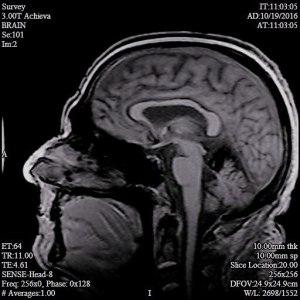
Gary started with a new group of doctors in 2017 and their first MRI included 436 images of his head and 85 of his spine. 14 months have passed so we asked if they would order an updated imaging set so we might see if there has been any reduction in the iron.
In the hopes we might learn something this time Gary asked Dr. Levy for his recommendation for the best MRI setting. It turns out the series from 2017 had a set with those very settings.
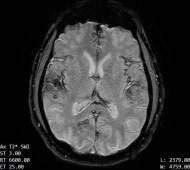
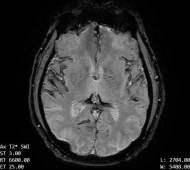
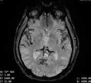
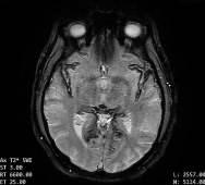
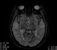
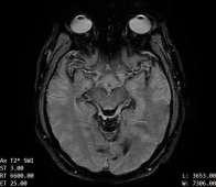
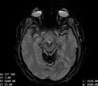
”The contours of the cerebrum, cerebellum, and brainstem are outlined with a T2 hypointense signal with blooming on susceptibility weighted sequences, which is compatible with the clinical history of superficial siderosis. A majority of the T2 hypointense signal is present in the superior folia of the cerebellum but also seen coating the surfaces of the brainstem, the cortical surfaces along the Sylvian fissures, and the cortical surfaces of the paramedian sulci of the frontal and occipital lobes. Few subcortical and periventricular T2/FLAIR hyperintensities are present in both cerebral hemispheres.”
If you look at each of the images you can clearly see the dark areas where his hemosiderin deposits are. The same machine and settings will be used so we should be able to visually compare the new images with these for some positive change.

