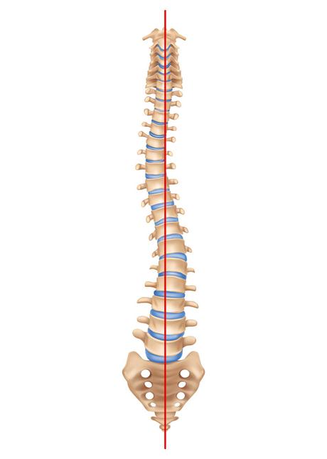
Scoliosis is an abnormal lateral curvature of the spine. It is often diagnosed in childhood or adolescence. Normal curves of the spine occur in the cervical, chest, and lower back parts on a plate called “sagittal”. These natural curves place the head on the pelvis and work as shock absorbers to distribute the mechanical pressure of the body as it moves. Scoliosis is often defined in the frontal coronal as the curvature of the spine. While the degree of bending is measured in the coronal plane, scoliosis is a more complex, three-dimensional problem that involves the following screens:
-
Coronal plate “Coronal”
Sagittal plate “Sagittal”
Pivot plate “Axial”
Prevalence and Spread of Scoliosis
Scoliosis affects 2-3% of the population or six to nine million people in the United States. Scoliosis can occur in the early stages of childhood. However, the early onset age of scoliosis is 10-15 years old and occurs in both sexes alike. In women, it progresses eight times more than usual, which requires treatment. Every year, scoliosis patients have more than 600,000 visits to a private doctor’s office, an estimated 30,000 children have “brace” bras, and 38,000 patients undergo spinal “fusion” fusion surgery.
Causes of Scoliosis or Spinal Deviation
Scoliosis can be classified according to the cause of the disease:
Idiopathic, congenial, or neuromuscular.
- Idiopathic scoliosis is diagnosed when all other causes of the disease have been ruled out. This type of disease accounts for about 80% of all cases. Idiopathic adult scoliosis is the most common type of scoliosis and is usually diagnosed during puberty.
- Congenital scoliosis “Congenital” is caused by a fetal malformation of one or more vertebrae and may develop anywhere in the spine. Vertebral abnormalities cause curvature of other parts of the body and spinal abnormalities, as one area of the spine grows at a slower rate than the rest. The size and location of abnormalities and child development determine the rate of scoliosis progression in adulthood. Since these abnormalities are present at birth, congenital scoliosis is usually diagnosed at a younger age than idiopathic scoliosis.
- Neuromuscular neuroplastic scoliosis involves secondary scoliosis that occurs in neuromuscular diseases. This type of scoliosis occurs in connection with cerebral palsy diseases, spinal trauma, muscular dystrophy, spinal muscular atrophy, and spina bifida “spina bifida”. In general, this type of scoliosis progresses more rapidly than idiopathic scoliosis and often requires surgical treatment.
Treatment of Scoliosis or Spinal Deviation
Once the diagnosis of scoliosis has been confirmed, there are several issues to assess that can help determine treatment options:
- Spinal maturation. Is the patient’s spine still growing and changing?
- The amount and degree of curvature. How severe is the curve and how can it affect a patient’s lifestyle?
- Curved location. According to some experts and with high probability, chest curves progress relative to those of other areas of the spine.
- The possibility of progressing the curve. Patients with large curvature before completing their age development are likely to experience curve progression.
After evaluating these variables, the following treatment options are recommended:
Care and observance of disease considerations
Bracing or Bracing
Surgery

In many children with scoliosis, the spinal curve is mild enough that it does not require treatment, but if the doctor is concerned that the curve will increase, it may examine the child every four or six months until they reach adulthood.
In adults with scoliosis, X-ray checking and treatment are usually recommended every 5 years unless symptoms gradually worsen.
Physiotherapy Of Shoulder Impingement Syndrome what is Hallux Valgus or Big Toe Deviation?Symptoms and Causes of Scoliosis or Spinal Deviation
Several symptoms may indicate scoliosis. If one or more of the following symptoms are taken into consideration by you and those around you, see your doctor.
- The shoulders are not smooth. One or both shoulder blades may have come out.
- The head is not directly above the pelvis.
- One or both sides of the hips and thighs are higher or placed in an unusual shape.
- The chest or ribs are in different places. (They are not consistent.)
- The waist and middle of the trunk are lumpy.
- Deformities the appearance or texture of the skin that covers the spine (bumps, hair spots, abnormal colour)
- The whole body leans to one side.
Idiopathic scoliosis may affect pulmonary function due to changes in the shape and size of the chest. Recent reports on pulmonary function testing in patients with mild to moderate idiopathic scoliosis show decreased and poor pulmonary function.
Diagnosis of Scoliosis
Scoliosis is usually confirmed through physical examination, X-ray, spinal radiography, CT scan, or MRI. The curve is measured using the Cobb method and is diagnosed by the degrees of measurement in terms of disease severity. The positive diagnosis of scoliosis is given based on the curvature of the measured coronal (if it is more than 10 degrees) in posterior-anterior radiography. In general, if it is more than 25 to 30 degrees, it is considered a significant curve. Curves above 45 to 50 degrees are considered severe and often require invasive and rapid treatment.
- A standardized and common exam, sometimes used by pediatricians and in school testing and screening, is called the “forward bending test.” During this experiment, the patient bends over each other by sticking his legs together, creating a 90-degree angle in his lower back. From this angle, any asymmetry of the trunk or any abnormal curvature of the spine is easily detected by your doctor. This is a preliminary screening test that can detect possible problems but cannot determine exactly the type or severity of the anomaly. Radiographic tests are needed to accurately diagnose and test positive for scoliosis.
- X-ray “X-ray”: The use of radiation to produce film or image of part of the body can show the structure of the vertebrae and a glimpse of the joints. X-rays of the spine are obtained to look for other possible causes of pain, i.e., infections, fractures, abnormalities, etc.
- Computerized tomography (CT or CAT scan): Reads the diagnostic image created after X-rays. Shows the shape and size of the spinal canal, its contents, and the surrounding structure. Very good at visualizing and simulating bone structures.
- Magnetic resonance MRI: A diagnostic test that produces 3D images of body structures using powerful magnets and computer technology. This method can show the spinal cord, nerve roots, and surrounding areas as well as enlargement, degeneration of “degeneration” and deformation.
Bracing for The Treatment of Scoliosis
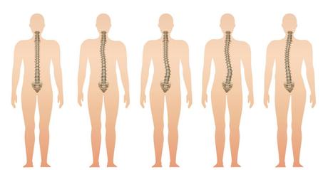
Braces are only effective in patients who have not reached skeletal maturity. If the baby is still growing and his curve is between 25 degrees and 40 degrees, a brace may be recommended to prevent the progression of curvature and bending. There have been improvements in brace design that, in newer models, braces are located under the arm, not on the neck. Different types of braces are available. While there is disagreement among experts over the choice of brace type, extensive studies show that braces reduce curve progression by about 80 percent if used and fully adhered to principles by children with scoliosis. For optimal effectiveness, braces should be checked regularly to ensure their suitability, it may also need to be worn for 16 to 23 hours a day to stop the growth.
Surgery to Treat Scoliosis
The two main objectives of surgery in children are to prevent curve progression in adulthood and reduce spinal abnormalities. Most specialists recommend surgery only if the curvature of the spine exceeds 40 degrees and there are signs of progression. Depending on the specific type of the disease, this surgery can be performed using the anterior method (through the front) or the posterior approach (through the back).
Some adults who are monitored as child treatment may need to have re-surgery, especially if they were treated 20 to 30 years ago. Spinal surgery should also be performed before making major improvements in the disease. In the past, a large part of the spine was fusion. When many parts of the vertebrae are intertwined, the remaining parts assume greater pressure from the load and stress caused by movements. This type of disease is a process in which degenerative changes occur in the upper and sub-fusion sections of the spinal cord. It can lead to painful arthritis of discs, facial joints, and ligaments.
