
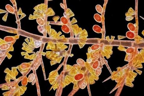
Arlene Wechezak snapped this image of red algae. The image shows its reproductive machinery. Red algae
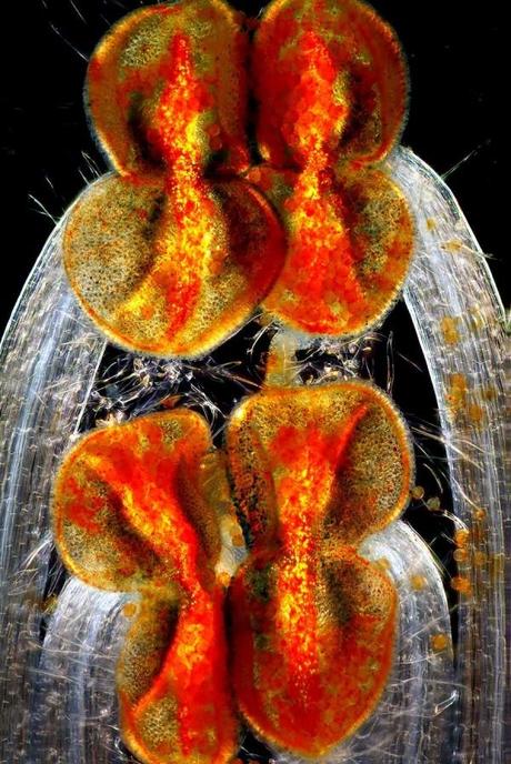
Edwin Lee got this image of the male reproductive parts of the Henbit, and annual plant that is something regarded as a weed. Henbit flower reproductive parts.
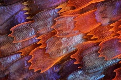
Charles Krebs shot the wings of a butterfly named “Prola Beauty” to capture these microscopic scales. Butterfly wing scales.
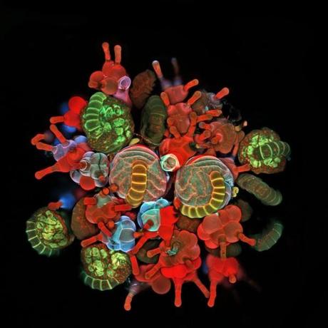
Igor Siwanowicz took this image of a common East-coast fern. It shows a cluster of spore-filled reproductive units and the specialized protective hairs around it. Fern spores.
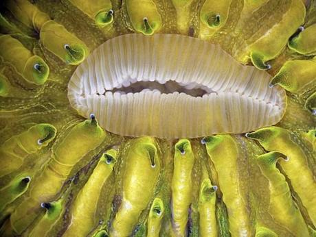
James Nicholson took this close-up of a live mushroom coral. Its mouth is in the process of expanding and the color comes from the coral glowing. Mushroom coral mouth
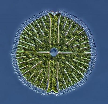
Rogelio Moreno Gill stacked 22 images to get this picture of a single-celled green algae from a lake. Green algae
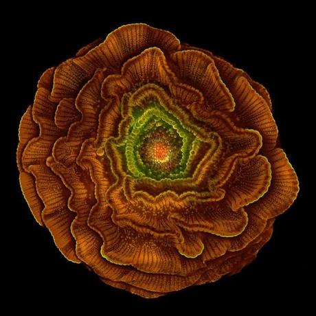
Sahar Khodaverdi got this image of the seed of the flowering plant Delphinium.
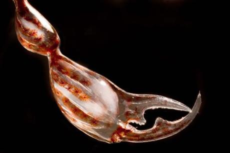
Christian Sardet and Sharif Mirshak took this picture of the claw of a crustacean that shows the muscles inside the claw and rows of pigment cells insider the shell.
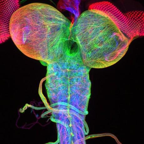
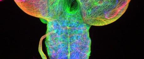
Christian Klambt and Imke Schmidt got this image of a fruit fly brain by marking the structural proteins of the cells. It shows the developing eye disks as well.
This incredible image of fern spores is just one of the many mind-blowing images that won the Olympus BioScapes Imaging Competition in 2012. Above are the top ten images and videos captured by life-science researchers.
The first prize winner, Ralph Grimm, took home $5,000 worth of Olympus equipment for his first-place video of tiny creatures known as rotifers.
“These fascinating and beautiful images tell important stories that shed light on the living universe around us, showing us the intimate structures and dynamic events of life in ways that we cannot ordinarily see,” Brad Burklow, of Olympus, said in a press release. “BioScapes movies and still images remind us of the fascination and beauty of the natural world, and highlight important work going on in laboratories across the globe.”
You can see all the runners up on the Olympus website. A selection of these images and movies will be displayed on a museum tour sometime this year.
via businessinsider
