Highlights
•In vivo Ca 2+ imaging of ∼12,000 neurons in mouse cortex at single-cell resolution
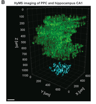
•Simultaneous 2p and 3p Ca 2+ imaging within 1,000 × 1,000 × 1,220 μm at up to 17 Hz
•Volumetric 3p Ca 2+ imaging of hippocampus through intact cortex
•A new integrated, systems-wide optimized microscopy design paradigmSummary
Calcium imaging using two-photon scanning microscopy has become an essential tool in neuroscience. However, in its typical implementation, the tradeoffs between fields of view, acquisition speeds, and depth restrictions in scattering brain tissue pose severe limitations. Here, using an integrated systems-wide optimization approach combined with multiple technical innovations, we introduce a new design paradigm for optical microscopy based on maximizing biological information while maintaining the fidelity of obtained neuron signals. Our modular design utilizes hybrid multi-photon acquisition and allows volumetric recording of neuroactivity at single-cell resolution within up to 1 × 1 × 1.22 mm volumes at up to 17 Hz in awake behaving mice. We establish the capabilities and potential of the different configurations of our imaging system at depth and across brain regions by applying it to in vivo recording of up to 12,000 neurons in mouse auditory cortex, posterior parietal cortex, and hippocampus.


