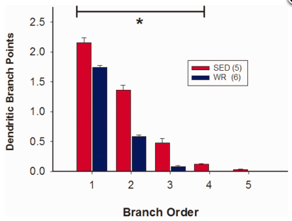
Here is yet another sobering note for couch potatoes. Lack of exercise (in rats) causes undesirable remodeling of the brain. Gretchen Reynolds points to work by Mischel et al. (open source) showing that inactive versus active rats show changes in the region of the rostral ventrolateral medulla that regulates the sympathetic nervous system, increasing connectivity and reactivity, potentially overstimulating the sympathetic nervous system to constrict blood vessels, increase blood pressure, and thus enhance the possibility of cardiovascular disease. Here is the Mischel et al. abstract and a summary figure:
Increased activity of the sympathetic nervous system is thought to play a role in the development and progression of cardiovascular disease. Recent work has shown that physical inactivity versus activity alters neuronal structure in brain regions associated with cardiovascular regulation. Our physiological studies suggest that neurons in the rostral ventrolateral medulla (RVLM) are more responsive to excitation in sedentary versus physically active animals. We hypothesized that enhanced functional responses in the RVLM may be due, in part, to changes in the structure of RVLM neurons that control sympathetic activity. We used retrograde tracing and immunohistochemistry for tyrosine hydroxylase (TH) to identify bulbospinal catecholaminergic (C1) neurons in sedentary and active rats after chronic voluntary wheel-running exercise. We then digitally reconstructed their cell bodies and dendrites at different rostrocaudal levels. The dendritic arbors of spinally projecting TH neurons from sedentary rats were more branched than those of physically active rats (P < 0.05). In sedentary rats, dendritic branching was greater in more rostral versus more caudal bulbospinal C1 neurons, whereas, in physically active rats, dendritic branching was consistent throughout the RVLM. In contrast, cell body size and the number of primary dendrites did not differ between active and inactive animals. We suggest that these structural changes provide an anatomical underpinning for the functional differences observed in our in vivo studies. These inactivity-related structural and functional changes may enhance the overall sensitivity of RVLM neurons to excitatory stimuli and contribute to an increased risk of cardiovascular disease in sedentary individuals.

Physical inactivity versus activity is associated with functional changes in control of blood pressure by neurons in the rostral ventrolateral medulla (RVLM). The present study shows that putative cardiovascular RVLM neurons have more complex dendrites in inactive versus active rats. This anatomical difference may underpin the functional differences previously reported.

