Professor Charles M Lieber says that the current methods we have for monitoring or interacting with living systems are limited. We can use electrodes to measure activity in cells or tissue, but any electrical probing tends to damage the cells.
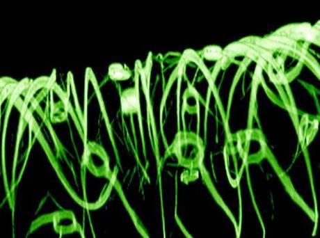
But recently, a research team led by Charles M. Lieber, the Mark Hyman Jr. Professor of Chemistry at Harvard, and Daniel Kohane, a Harvard Medical School professor in the Department of Anesthesia at Children’s Hospital Boston, developed a system for creating a type of “cyborg” tissue for the first time by embedding a three-dimensional network of functional, biocompatible, nanoscale wires into engineered human tissues.
In a process similar to making microchips, the wires and a surrounding organic mesh are etched onto a substrate, which is then dissolved, leaving a flexible mesh. Groups of those meshes are formed into a 3D shape, then seeded with cell cultures, which grow to fill in the lattice to create the final system. Scientists were able to detect signals from heart and nerve cell electro-flesh made this way, allowing them to measure changes in response to certain drugs. In the near-term, that could allow pharmaceutical researchers to better study drug interaction, and one day such tissue might be implanted in a live person, allowing treatment or diagnosis.
Or allowing him/her to become a cyborg/bionic.
N.


Image: 3D reconstruction confocal microscopy image of a 3D macroporous nanoelectronic scaffold; a nanoelectronic device exists (too small to see in image) in each circular region.


